Background: How do fossils preserve?
Unaltered hardparts
The concept of unaltered remains can refer to multiple modes of preservation. Freezing, encapsulation in amber (tree resin), desiccation, and chemical preservation, such as entombment in petroleum containing sediment, are various examples. Freezing, mummification (desiccation), oil seeps, and amber can preserve both soft and hard tissues. Sometimes the soft tissues decay, but the hard parts remain unaltered. Teeth, bones, and shells may be preserved as unaltered hard parts if the chemical environment allows it. The specimen below is an ammonite that preserves its original shell nacre (‘mother of pearl’)- it had been preserved in an oil seep.
Click here to see a full screen 3D model of the ammonite.
Replacement
Replacement occurs when the pore spaces of organic tissues are impregnated with dissolved minerals. Common minerals found in permineralised fossils include silica, calcite, phosphates, pyrite, iron oxides.
The example below is a brachiopod that has been replaced by silica. The original shell of organisms like brachiopods and bivalves are made of calcium carbonate. When such organisms undergo replacement in the fossil record, they commonly have a grey colour, and are generally much more resilient to erosion/destruction than the original skeleton. Bones of vertebrate animals (e.g., dinosaurs) commonly become fossilised this way too).
Section 1: Styles of fossil preservation (external students only)
Fossil 1: Brachiopod (Permian)
Click here to see a full screen 3D model of this specimen.
Fossil 2: Oyster (Carboniferous)
Click here to see a full screen 3D model of this specimen.
Fossil 3: Tree (Cenozoic)
Click here to see a full screen 3D model of this specimen.
Fossil 4: Elephant tooth (Pleistocene)
Click here to see a full screen 3D model of this specimen.
Fossil 5: Seed fern (Triassic)
Click here to see a full screen 3D model of this specimen.
Fossil 6: Ammonite (Cretaceous)
Click here to see a full screen 3D model of this specimen.
Fossil 7: Seed fern (Triassic)
Click here to see a full screen 3D model of this specimen.
Fossil 8: Coral (Carboniferous)
Click here to see a full screen 3D model of this specimen.
Fossil 9: Baragwanathia (plant; Silurian)
Click here to see a full screen 3D model of this specimen.
Fossil 10: Ammonite (Cretaceous)
Click here to see a full screen 3D model of this specimen.
Section 2: The who’s who of the dinosaur family tree
If you’re an iPhone or iPad user, download the ‘Dinosaur Discovery’ app here to complete this exercise. To access the dinosaur tracker codes, click here. These trackers will work best if you print them as a hardcopy and then cut them out with scissors.
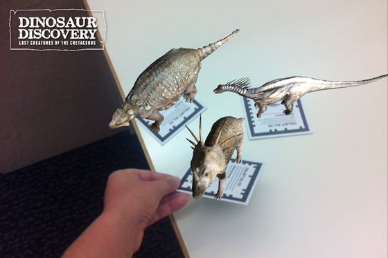
Unfortunately this app is no longer supported for Android or other non-Apple devices. To remedy this, if you cannot access the app please see the below video of Kev’s clumsy thumbs showing you the various dinosaurs.
Dinosaurs
Section 3: Skull parade
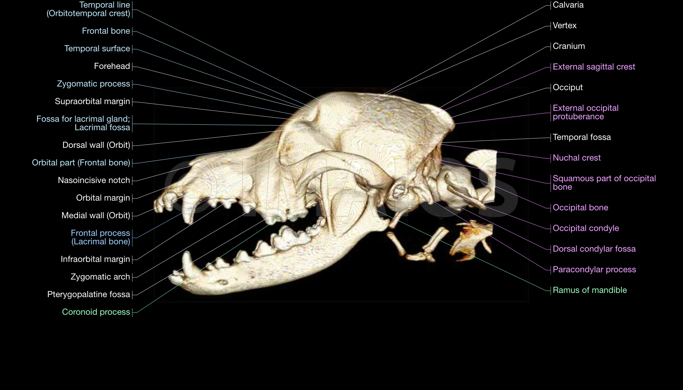
See below for the 10 different marsupial and placental skulls, randomly divided into two sets. If you wish to use correct anatomical terms to describe the morphological differences that you think are useful, feel free to use this guide (click to enlarge):
Mammal skull set 1:
Click here to see a full screen 3D models of Set 1.
Mammal skull set 2:
Click here to see a full screen 3D models of Set 2.
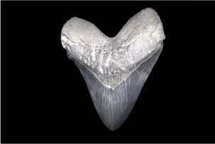
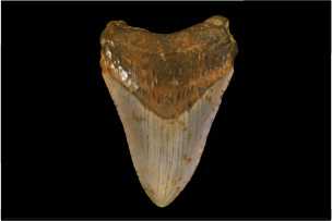
Section 4: How big was megalodon?
This is a reconstruction of megalodon. The largest individuals are thought to have reached up to 18 m although animals of this size were very rare.
Megalodon tooth 1: click thumbnail image below to open the 3D model and measuring tool. To measure the tooth, after the model has loaded, click the ruler to the upper left and follow the instructions from there. The key measurement to take on each tooth is the ‘major slant height’ (see exercise sheet for details).
Megalodon tooth 2: click thumbnail image below to open the 3D model and measuring tool.